Full-Field Digital Screening and Diagnostic Mammography

Mammography Screening has been shown to reduce breast cancer mortality and save lives. The American College of Radiology recommends mammography screening annually for women beginning at age 40. A baseline mammogram may be obtained between ages 35-40.
An abnormal screening mammogram may lead to a diagnostic mammogram, in which a specific abnormality needs to be further evaluated with additional images. Diagnostic mammography may also be performed on patients with clinical or physical signs of breast disease.
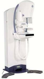
Digital mammography is beneficial in that it utilizes less radiation in image acquisition and provides improved image quality, as compared with film-screen mammography. Digital mammography has been shown to be especially helpful in evaluating patients with dense breast tissue, due to improved image accuracy. At J Gershon Breast Imaging, all mammograms are obtained on a 2013 GE Senographe Full-Field Digital Unit.
What about 3D mammography?
3D mammography, also known as breast digital tomosynthesis, is an emerging technology for breast imaging. The imaging is performed by obtaining multiple images of the breast in an “arc like” fashion. This allows the radiologist to view the breast in multiple “slices”, as opposed to the standard two views (2D images) of each breast.
3D mammography is being marketed as having increased sensitivity in detecting cancers. However, at this time, it has only been found to detect approximately 1-2 additional cancers per 1000. In fact, an article published in the American Journal of Roentgenology, January 2019, found no difference in the cancer detection rate between 2D and 3D digital mammography. The main advantage of 3D imaging is that is decreases call-back rates for the radiologist. In other words, less women have to return for additional imaging if a finding is seen on the mammogram. 3D imaging is most useful in women with dense breast tissue. However, recent studies show that ultrasound is a more sensitive tool for detecting cancers in dense breast patients.
3D imaging has its disadvantages. In many situations, 2D imaging must still be obtained first, followed by 3D imaging. This essentially doubles the radiation dose to the patient.
At J Gershon Breast Imaging, it is our belief that a top quality low dose 2D digital mammogram, interpreted by an experienced breast radiologist, is equivalent to, if not better than, a 3D study. At our office we do not have “call-backs”, as every study is interpreted immediately. Additionally, we have found that screening breast ultrasounds for dense tissue, which impose no additional radiation dose, are highly sensitive for detecting small invasive cancers not visible in dense breast tissue, both on 2D and 3D imaging.