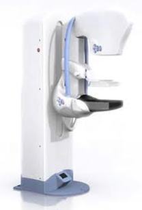
 Our current society is one of a “digital” age. Almost everything we do on a day-to-day basis involves some form of digital media. Over the years radiology imaging has joined the digital era and “films” have become a word of the past. The first full-field digital mammography unit was approved by the FDA in 2000. Since that time, the number of facilities converting to full-field digital mammography has multiplied rapidly.
There are two types of digital mammography; full-field digital mammography (FFDM) and computed radiography (CR). CR units were introduced as a less-expensive alternative to FFDM, as offices could modify older screen-film mammography equipment and convert the images into a digital format. However, it has been found that the cancer detection rates are lower with CR vs. FFDM. CR was found to be 21% less effective than FFDM in detecting cancer.(1) A recent comparative study found that FFDM has significantly better image quality with regards to tissue coverage, compression, exposure, contrast, and resolution.(2) Women with dense breast tissue benefit significantly from FFDM, as the tissue is more clearly visualized and calcifications are more sharply delineated. In addition to improved quality and better cancer detection with FFDM, there is less radiation per image, 22%, when compared to film-screen mammography.(3) When choosing a mammography center, it is important to know exactly what type of mammogram you will be undergoing. There is “digital” imaging and there is “full-field” digital imaging. Be sure to ask what type of mammogram you will be receiving and choose your mammography center wisely. As the benefits of FFDM outweigh CR, why sacrifice your care when you can have the best there is to offer?! Julie S. Gershon, M.D. Breast Imaging Specialist (1) "Digital Compared with Screen-Film Mammography: Performance Measures in Concurrent Cohorts within an Organized Breast Screening Program." Collaborating with Dr. Chiarelli were Sarah A. Edwards, M.H.Sc., Maegan V. Prummel, M.P.H., Derek Muradali, M.D., Vicky Majpruz, M.Sc., Susan J. Done, M.B., B.Chir., Patrick Brown, Ph.D., Rene S. Shumak, M.D., and Martin J. Yaffe, Ph.D. The study was funded by the Canadian Institutes of Health Research-Radiology May 2013 (2) "A Comparative Study of Computed Radiography-based Mammography Using Digital Phosphor Storage Plate and Full Field Digital Mammography” Chelliah KK, Tamanang S, Bt Elias LS, Ying KY - Indian J Med Sci. 13 Oct. 2013. (3) "Comparison of Acquisition Parameters and Breast Dose in Digital Mammography and Screen-Film Mammography in the American College of Radiology Imaging Network Digital Mammographic Imaging Screening Trial” R. Edward Hendrick, Etta D. Pisano, Alice Averbukh, Catherine Moran, Eric A. Berns , Martin J. Yaffe, Benjamin Herman, Suddhasatta Acharyya and Constantine Gatsonis-American Journal of Roentgenology: Vol. 194, No. 2 (AJR) Feb. 2010.
2 Comments
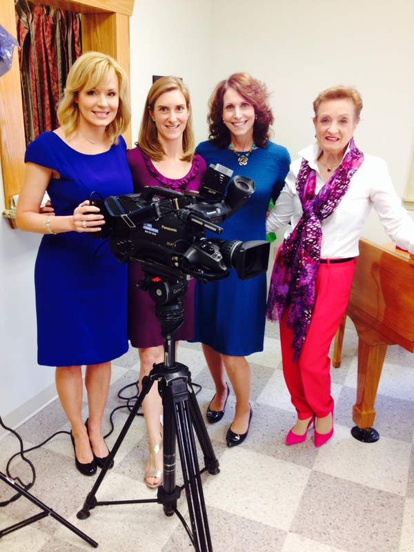 Lisa, Julie, Nancy, & Jan Lisa, Julie, Nancy, & Jan We were welcomed yesterday with a visit from Lisa Carberg of NBC Connecticut. Lisa and crew came to the office to film a story on dense breast tissue. I thank Lisa and my "dense breast buddies", Nancy Cappello and Jan Kritzman, for helping to make this event possible and for the ability to share this important information with other women.
Are you aware that in the state of Connecticut you are required to be notified of your breast density? In 2009, Connecticut was the first state to pass notification legislation and there are currently only 13 other states with this legislation in place. What does it mean to have dense breasts? When the majority of the breast is composed of fibrous and glandular tissue, a woman is reported to have "dense breasts". While dense breast tissue is more common in younger women, it is also seen in older women. Approximately half of all women have dense breast tissue. What is the concern? Women with dense breast tissue have a greater risk of breast cancer. Interpretation of dense breast mammograms can be markedly limited depending on how much dense tissue is present. Cancers can hide in dense tissue and may not be detected on a mammogram. What is recommended for dense breasts? Routine screening mammograms should be obtained. A screening breast ultrasound is also highly recommended, as this gives the radiologist a better look into the dense tissue. Screening breast ultrasound has been shown to detect cancers not visible on the mammogram and can be a life saving study. Watch for our upcoming news story and feel free to contact the office with any questions or concerns regarding breast density! |
AuthorJulie S. Gershon, M.D. Archives
October 2023
Categories |

 RSS Feed
|
J Gershon Breast Imaging
|
21 Arch Rd. Avon, CT 06001
|
P: 860.673.8379 F: 860.271.8025
|