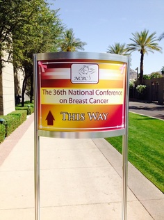
 For those who have heard of 3D mammography, and those who have not yet, I would like to share some information I learned at the American College of Radiology’s National Conference on Breast Cancer. 3D mammography, also known as digital breast tomosynthesis, is a relatively new technology, however it is currently not FDA approved as an alternative to standard 2D mammography. The 3D mammogram is obtained with an x-ray unit that moves in an “arc” around the breast taking multiple images which are then reconstructed by a computer into a 3D image. The goal of the technology is to allow the radiologist a clearer visualization the breast tissue at multiple levels with the hope of improving mammographic interpretations. The main advantage of breast tomosynthesis is that it reduces patient call-backs, with the hope of decreasing patient anxiety. Patients benefiting most from 3D imaging are those with dense breast tissue, as this is where the abnormalities hide. However, more is not always better, and there are many reasons why I am not fully supportive of breast tomosynthesis. Firstly, every patient that undergoes a 3D mammogram also needs a 2D mammogram, so patient radiation dose is doubled. Secondly, calcifications are are not well seen on 3D images, as they become blurred in the imaging process. Thirdly, in dense breast patients, ultrasound is still commonly needed as an additional study to further evaluate a 3D finding. And finally, insurance is currently not paying for breast tomosynthesis. While digital breast tomosynthesis may be of use in certain cases, I do not see an advantage for its use routinely at this time. With regards to decreasing patient anxiety, I feel the best way to remedy this issue is to speak to the patient and discuss the mammographic results at the time of exam. And for patients with increased breast density, we are fortunate to live in a state where breast density is acknowledged and where insurance will pay for a screening breast ultrasound. In dense breast patients, a full-field digital 2D mammogram and bilateral breast ultrasound is the best way to screen for breast cancer, and there is no added radiation! When choosing a breast imaging center, it is important to be aware of the various types of mammography being offered and to make an educated decision about the type of care you wish to receive. Julie S. Gershon, M.D.
0 Comments
Leave a Reply. |
AuthorJulie S. Gershon, M.D. Archives
October 2023
Categories |
|
J Gershon Breast Imaging
|
21 Arch Rd. Avon, CT 06001
|
P: 860.673.8379 F: 860.271.8025
|

 RSS Feed